การเปรียบเทียบคุณภาพภาพรังสีจากแผ่นรับภาพรังสีดิจิทัลในปากและเครื่องแปลงสัญญาณภาพรังสีเป็นระบบดิจิทัล
Comparison of radiographic image quality between intraoral digital imaging plates and digital radiography scanners
Keywords:
Radiographic image quality / Intraoral digital imaging plate / Digital radiography scanner / Dental radiography / Dental radiologyAbstract
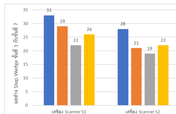
การถ่ายภาพรังสีในช่องปากระบบดิจิทัลแบบทางอ้อมมีความจำเป็นต้องใช้แผ่นรับภาพรังสีดิจิทัลในปากร่วมกับเครื่องแปลงสัญญาณภาพรังสีเป็นระบบดิจิทัล ซึ่งความแตกต่างของชนิดและยี่ห้อของแผ่นรับภาพรังสีดิจิทัลในปากและเครื่องแปลงสัญญาณภาพรังสีเป็นระบบดิจิทัลอาจส่งผลต่อคุณภาพของภาพรังสีที่ใช้ในการวินิจฉัยทางทันตกรรม การศึกษานี้มีวัตถุประสงค์เพื่อเปรียบเทียบคุณภาพของภาพรังสีที่ได้จากแผ่นรับภาพรังสีดิจิทัลในปาก 4 ยี่ห้อ และเครื่องแปลงสัญญาณภาพรังสีเป็นระบบดิจิทัล 3 ยี่ห้อ โดยการจัดวางแผ่นรับภาพรังสีดิจิทัลในปากคู่กับ Step wedge ใส่ในอุปกรณ์ยึดจับแผ่นรับภาพรังสี (XCP holder) โดยถ่ายภาพรังสีด้วยเครื่องเอกซเรย์และหุ่นจำลองเดียวกัน ค่าพารามิเตอร์เท่ากัน (70 kV, 6 mA, 0.25 sec) จากนั้นนำภาพรังสีที่ได้ไปสแกนด้วยเครื่องแปลงสัญญาณภาพรังสีเป็นระบบดิจิทัลแต่ละยี่ห้อ และวิเคราะห์ค่าความเข้มของเฉดสีด้วยโปรแกรมวิเคราะห์ภาพ ImageJ เพื่อประเมินช่วงความกว้างของระดับเฉดสี นอกจากนี้ยังประเมินจำนวนขั้นของ step wedge ที่สามารถมองเห็นได้ด้วยตาเปล่า ผลการศึกษาพบว่า แผ่นรับภาพรังสีดิจิทัลในปากยี่ห้อ P1 มีแนวโน้มให้คุณภาพภาพรังสีที่ดีกว่ายี่ห้ออื่น ในขณะที่ยี่ห้อ P3 มีแนวโน้มให้คุณภาพที่ลดลง สำหรับเครื่องแปลงสัญญาณภาพรังสีเป็นระบบดิจิทัล พบว่า ยี่ห้อ S1 มีแนวโน้มแสดงช่วงระดับเฉดสีได้กว้างและสามารถแยกขั้นของ step wedge ได้มากกว่า ในขณะที่ยี่ห้อ S3 มีแนวโน้มให้ผลที่ลดลง ดังนั้นชนิดของแผ่นรับภาพรังสีดิจิทัลในปากและเครื่องแปลงสัญญาณภาพรังสีเป็นระบบดิจิทัล แสดงให้เห็นถึงแนวโน้มความแตกต่างของค่าช่วงระดับเฉดสีระหว่างยี่ห้อ ผลการศึกษานี้สามารถใช้เป็นข้อมูลเบื้องต้นสนับสนุนการตัดสินใจในการจัดซื้อและทดแทนอุปกรณ์แผ่นรับภาพรังสีดิจิทัลในปากและเครื่องแปลงสัญญาณภาพรังสีเป็นระบบดิจิทัล เพื่อเพิ่มความแม่นยำและประสิทธิภาพในการวินิจฉัยและวางแผนการรักษาทางทันตกรรม
Indirect digital intraoral radiography requires the use of intraoral digital imaging plates in combination with digital radiography scanner systems to convert X-ray signals into digital images. Variations in the type and brand of intraoral digital imaging plates and scanners may affect radiographic image quality, which is essential for accurate dental diagnosis. The aim of this study was to compare radiographic image quality obtained from four brands of intraoral digital imaging plates and three brands of scanners. Each imaging plate was positioned in contact with an aluminum step wedge and secured in an XCP holder. All radiographic exposures were performed using the same intraoral X-ray unit and phantom under identical exposure parameters (70 kV, 6 mA, 0.25 s) to control for geometric and exposure-related variables. After exposure, the imaging plates were digitized using each scanner system. Quantitative image analysis was conducted using ImageJ software to evaluate grayscale intensity values and to assess the grayscale dynamic range. In addition, visual image quality was assessed by counting the number of step wedge levels that could be visually distinguished. The results demonstrated that intraoral digital imaging plate brand P1 provided the highest radiographic image quality, whereas brand P3 showed the lowest performance. Among the scanners, brand S1 exhibited the widest grayscale dynamic range and allowed visualization of the greatest number of step wedge levels, while brand S3 showed the poorest performance. In conclusion, the type of intraoral digital imaging plate and scanner system significantly influences radiographic image quality. These findings provide evidence to support informed decision-making regarding the procurement and replacement of intraoral digital imaging plates and scanners, thereby improving the accuracy and efficiency of dental diagnosis and treatment planning.